Strip micrograft or FUT is a minimally invasive technique that involves extraction of a strip of scalp from the occipital region. Micrografts with 1 to 4 hair follicles are obtained and these are implanted in the recipient site.
The strip hair graft technique is performed in three stages:

In most cases, the patient is advised to take a muscle relaxant or anxiolytic to reduced discomfort during the 4 to 6 hours of surgery.
If the patient is concerned with pain and anxious, a mild sedative can be administered, monitored by an anaesthetist. In this case, the technique must be performed in an IML operating theatre, equipped with the monitoring systems required for sedo-analgesia.
The donor site is the occipital region, although in some cases the temporal region can be used.
The occipital region is the area with most hair and less tendency to suffer from androgenic alopecia.
The donor site is typically 5 cm wide and approximately 25 cm long in the area between the ears. The strip should not be extracted in a high area, since it could be affected over time by progressive hair loss on the crown.
The size of the strip depends on the follicular unit density on the patient's scalp in this area.
The strip should contain the square centimetres needed to obtain the number of grafts agreed on with the patient for the transplant.
The strip width should not be more than 1 cm so the suture can be performed with no tension on the donor site, which ensures a good quality scar. Therefore, strip size is adapted, making it longer or shorter, according to the number of grafts required.
Once the length and width of the strip is decided, local anaesthetic is administered. A 1% lidocaine solution is infiltrated with adrenalin 1:1,000,000 diluted in 20 or 30 ml saline solution. This provides a good level of anaesthesia, vasoconstriction and turgidity in the infiltrated area, which will enable the strip extraction manoeuvre.
The strip is extracted using a fine-blade scalpel. The scalpel is placed in the direction of hair growth on the occipital region for several reasons:
The closure on the donor site is performed with a direct suture, with loose stitches or staples, assuring that the edges of the trichophytic incision match and they are close together to prevent the scar from broadening.

Follicular units are kept moist until the implantation takes place
The extracted strip is fragmented in follicular units with 1, 2 or 3 hairs, so they can be grafted onto the recipient site.
This painstaking task is performed using magnifying glasses, which improve precision when cutting and fragmenting the strip.
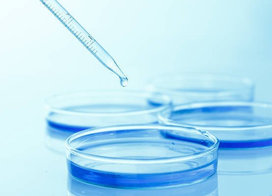
The strip is divided into narrower, lengthwise strips and then each of these are fragmented transversally into follicular units. Given that the resulting follicular units have 1 to 3 hairs, they are grouped according to the number of hairs they contain, placing them on gauze or Petri dishes.
Follicular units are kept moist in a trough with saline solution to assure vitality and viability when grafting.
Selection of the follicular units by the number of hairs they contain will aid the surgeon later. It is preferable to graft the follicular units with just one hair following the new hairline for a natural result. Grafts with more follicles are placed further back.

The incisions where each micrograft will be placed are first made. The following aspects must be taken into consideration:
The implantation line must be at least 8 cm from the glabella (between the brows). It should be an irregular contour and follow temporal hairline. These conditions are essential for a natural result.
The same type of solution as for the donor site is infiltrated, with ring block infiltration around the whole recipient site to improve anaesthesia.
Tumescent infiltration of the recipient site causes the tissues to distend and temporarily increase the skin surface. This enables placement of the grafts closer to each other.
The tumescent anaesthetic should not be too intense. If there is too much tension in the area the so-called "popping" effect occurs, where grafts are extruded by the excessive pressure.
Incisions are made with 20 G or 22 G needles for single hair grafts and 18 G for thicker grafts with 2 or 3 hairs.
The narrower incisions are made in the hairline where the single hair grafts are placed. The larger incisions are made towards the back where the 2 or 3 hair grafts are placed.
Incisions are made at an average depth of 5 mm to ensure correct graft reception, avoiding the risk of too deep an implantation which could cause folliculitis.
The distance between the incisions will determine the resulting grafted hair density. For a result to be natural, density should be 20 to 40 follicular units per square centimetre.
Orifice orientation is another essential factor for a natural result, this will later determine the growth direction of the grafts. Therefore, these incisions must follow the angle in which hair grows on its own.
Hair grows forward in the front, so the incisions are made at a 30º angle towards the front. As incisions are made further back, the orientation is gradually corrected until it reaches an angle of approximately 60º in the back.
The grafts are handled with fine-grade forceps, avoiding the lower part of the follicles and picking them up by the subcutaneous cell tissue that surrounds them.
They are clamped gently to prevent trauma or deformation and then implanted applying minimal pressure. This part of the surgery is highly dependent on technique and the surgeon's dexterity is of utmost importance.
Work is performed under magnifying glasses. This stage typically lasts 2 or 2.5 hours.
Would you like to know if the FUT technique is suitable for you? Request a free informative consultation now with one of our expert surgeons from our Hair Transplant Unit.